
Size of Kidney’s Glomerular in Children May Help to Diagnose Alport


Analyzing the appearance of the glomerular, using a method called glomerular morphometry, may help to diagnose Alport disease and other kidney conditions in children.
The study, “Contribution of glomerular morphometry to the diagnosis of pediatric nephropathies,” published in the journal Saudi Journal of Kidney Diseases and Transplantation, also suggested that linking such findings with laboratory data may aid in understanding how kidney disease progresses.
Since kidney disease in children rarely progresses quickly, physicians seldom do kidney biopsies in this patient group. Researchers at the Federal University of Triângulo Mineiro, in Brazil, argued that improving analysis of tissue using morphometry (measures of external form, or size and shape) may make it easier to diagnose kidney disease in pediatric patients.
They recruited 48 children and adolescents, ages 2-18, who underwent a kidney biopsy as part of a clinical evaluation. The children had a variety of diagnoses, including five with Alport syndrome.
As a comparison, the research team analyzed data from deceased individuals without kidney disease. In addition to morphometric analysis of the kidney, researchers analyzed protein levels in the urine, as well as urea and creatinine values. Creatinine is a waste product, often measured in the blood to detect kidney disease and assess kidney function.
The team found that the area of the glomerular capsule space (often called Bowman’s capsule) was larger in the control group than in patients with Alport disease or thin basement membrane disease. Another group of kidney disease patients, those having a condition called podocytopathy, also shared this feature.
Researchers also found that the Bowman’s capsule area correlated with the amount of urine protein, creatinine, and urea measured in the patients. Another measure — the glomerular tuft area — was also linked to the lab values of proteins, urea, and creatinine.
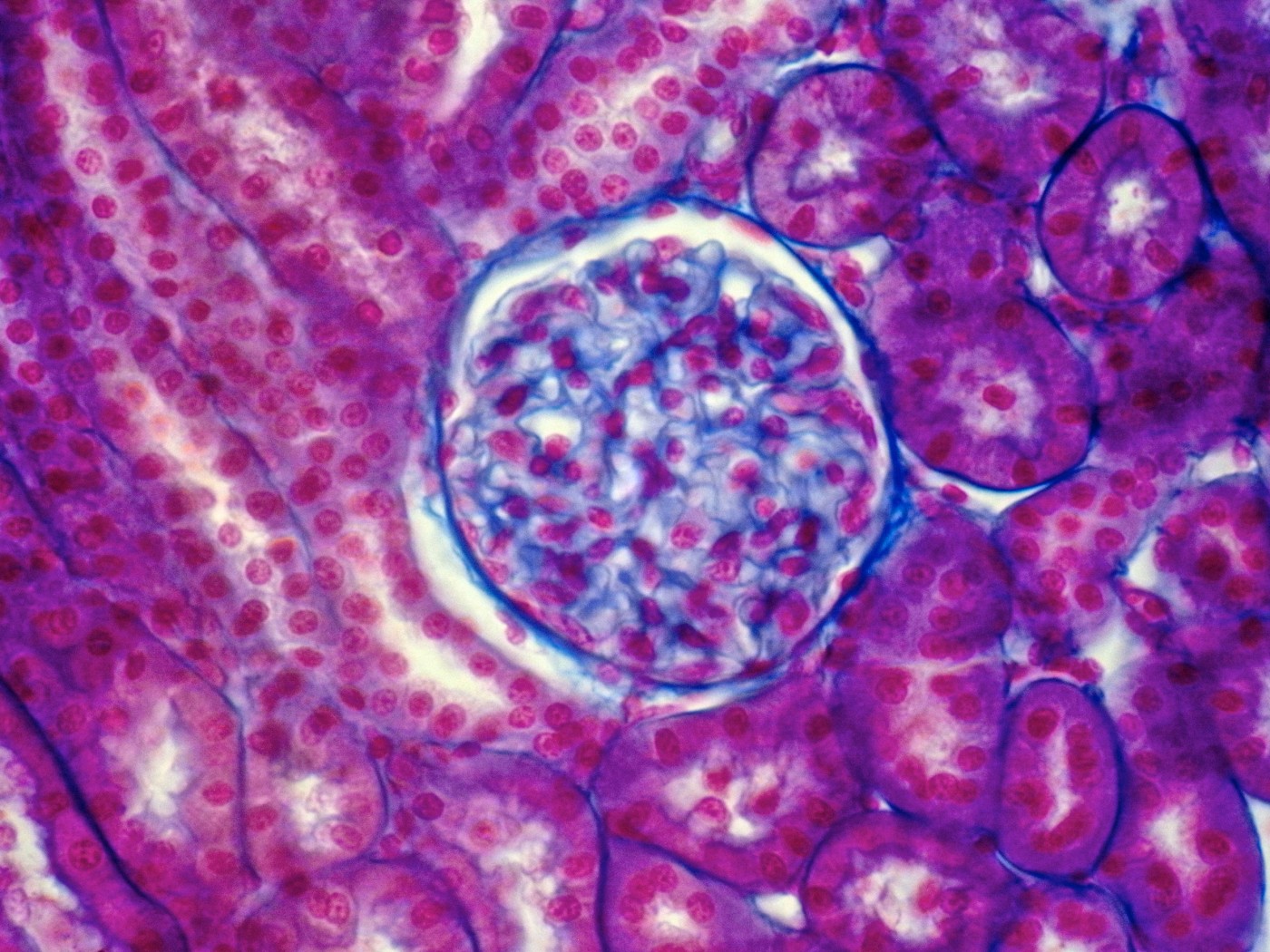
The glomerular tuft is the network of tiny blood vessels within the glomerulus, providing blood for filtration within the kidney. The links between glomerular anatomy and lab values held true in children with all types of kidney disease.
Team members argued that the small glomeruli of Alport patients, including the reduction of Bowman’s space, could be explained by changes known to occur in the kidneys because of the disease. “Furthermore, the association between glomerular morphometric parameters and laboratorial data can potentially promote a better understanding of the pathogenesis of kidney diseases, especially in pediatric patients in whom studies are less frequent,” they concluded.


Leave a comment
Fill in the required fields to post. Your email address will not be published.