Kidney Cells in Urine May Be a New Tool to Diagnose and Treat Alport, Researchers Say

A new method of analyzing the activity of COL4A genes in the kidney cells of patients with suspected or confirmed Alport syndrome may improve molecular diagnostics and prognostic assessments, say researchers at Italy’s University of Siena.
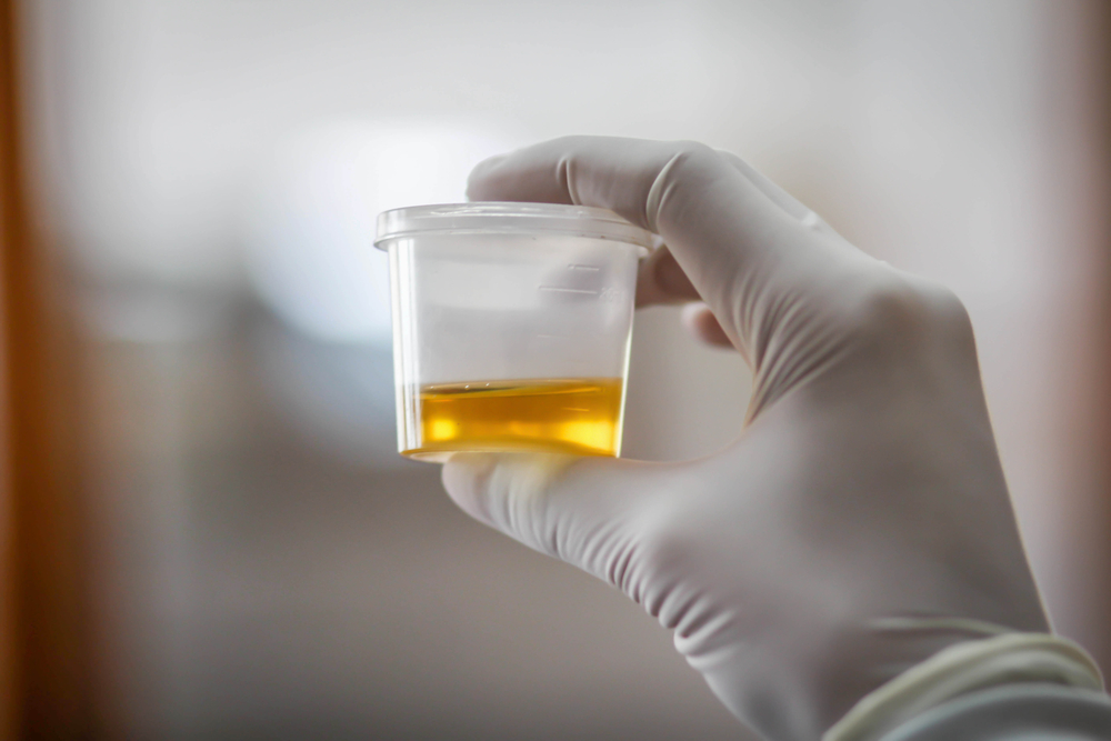
The team isolated podocyte cells — which make up the largest part of the kidney glomerular basement membrane, destroyed in Alport — from urine samples. This let them study how mutations directly affect gene activity in diseased cells without invasive kidney biopsies.
The study, “Urine-derived podocytes-lineage cells: a promising tool for precision medicine in Alport,” appeared in the journal Human Mutation. It concluded that an easily available source of diseased cells opens up the possibility of testing personalized gene therapies that target specific Alport mutations.
Podocytes are kidney cells that line the tiny blood vessels of the glomeruli to help filter blood into urine. These cells are the only known cells producing all three versions of COL4A that are mutated in various Alport types.
While these cells likely play a key role in Alport disease processes, they have been difficult to study, since non-invasive approaches to collect them have not been available. Studies in patients with other types of diseases destroying the kidney basement membrane, however, suggest it is possible to isolate these cells from a urine sample.
To test whether this is possible also in Alport, the team gathered urine samples from 25 patients. They managed to isolate podocyte kidney cells and grow them in lab dishes.
Analyses showed that the COL4A3, COL4A4, and COL4A5 genes were active in these cells. Researchers then assessed if and how certain mutations — which earlier research could not prove were causing disease — contributed to disease processes. They could confirm that five such mutations did cause Alport by affecting so-called splicing processes.
When a gene is translated into a protein, stretches of DNA, not coding for protein, need to be cut out and the remaining pieces stitched together. This process is termed splicing and it was disrupted by some of these mutations, leaving the proteins either truncated or faulty in other ways.
Besides providing researchers with a tool for studies of disease mechanisms and gene therapies, researchers said the cells offer an opportunity to assess the effects of other treatments now being tested in patients.


Leave a comment
Fill in the required fields to post. Your email address will not be published.