
Researchers Study Rare Case of a Foodpipe Abnormality in Boy with Alport Syndrome


Michigan researchers reported the rare case of a 7-year-old boy with a foodpipe abnormality stemming from his Alport syndrome.
About 5 percent of Alport patients develop a condition called diffuse leiomyomatosis. Its hallmarks are an abnormal increase in the number of smooth muscle cells that facilitate certain functions in the body, and weakness in those muscles.
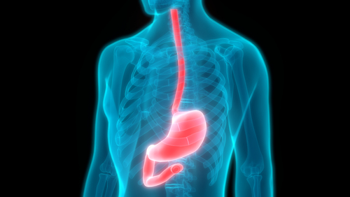
The condition mainly affects the foodpipe, or esophagus; the tracheobronchial wall, or windpipe wall; and women’s sex organs. The esophagus is the tube that carries food from the mouth to the stomach.
Scientists don’t fully understood what causes leiomyomatosis. But two-thirds of the esophagus-related cases occur in Alport syndrome patients, suggesting a link between the two.
Previous studies have suggested that the answer lies in mutations of the COL4A5 and COL4A6 genes. This research involved scientists analyzing esophagus tissue collected from Alport patients with diffuse leiomyomatosis. A key finding was that the COL4A5 and COL4A6 genes in the tissue were unable to generate protein.
The findings failed to shed light on the genes’ role in the abnormal increase in smooth muscle cells and in Alport patients’ development of leiomyomatosis.
Researchers at the Children’s Hospital of Michigan decided to study the case of a boy with Alport syndrome who had been experiencing vomiting episodes for several years.
Their article, “7-year-old with Alport Syndrome and Vomiting,” appeared in the journal Gastroenterology.
At the time of the boy’s first evaluation, he was vomiting daily, usually within an hour of eating. He had no additional symptoms, such as abdominal pain, chest pain, painful or difficult swallowing, or weight loss.
Further evaluations showed that he had an enlarged esophagus. In addition, his esophagus-related muscle tissue was thicker than normal, and thicker than that of other wall muscles in the chest.
The team concluded that he had diffuse esophageal leiomyomatosis.
His condition worsened to the point where he was unable to tolerate solid food — and then liquids.
When he was 10, doctors removed his esophagus. They replaced it with one constructed from stomach tissue in a procedure called reverse gastric tube esophageal replacement. This led to the boy being able to eat again.
“Accurate diagnosis is essential for these patients because treatments such as myotomy [cutting the muscle] and dilatation [stretching it] will not result in symptom resolution and can complicate surgical treatment,” the researchers concluded.


Leave a comment
Fill in the required fields to post. Your email address will not be published.