Sulfatides May Play Important Role in Early Stages of Alport Syndrome, Mouse Study Says

Scientists found two molecules called sulfatides in the kidneys of a mouse model of Alport syndrome that may play an important role during the early stages of the disease.
The findings of the study, “Two Specific Sulfatide Species Are Dysregulated during Renal Development in a Mouse Model of Alport Syndrome,” were published in Lipids.
Alport syndrome is a genetic disorder characterized by progressive kidney disease that can lead to kidney failure. It is caused by genetic mutations in either COL4A3, COL4A4 or COL4A5, which provide instructions to build different subunits of a large protein called type 4 collagen (COL4). COL4 is the main component of connective tissue.
Progressive kidney disease associated with Alport is caused by the gradual loss of function of the glomerular basement membrane (GBM), an important component of the kidney’s filtration system. The GBM is made up of COL4 and laminin, another protein that is one of the major components of basement membranes.
“Normally, laminin α1β1γ1 (LM-111) is expressed only during the early stages of glomerular development. In Alport syndrome, the LM-111 is reexpressed [produced again] in the adult GBM, although the mechanisms governing this re-expression are unclear,” the investigators said.
“It has been shown that sulfatides, a class of acidic glycosphingolipids [a subtype of fatty molecules that are part of the cell membranes], can initiate basement membrane assembly by binding directly to LM-111. This finding suggested that sulfatides within the plasma membrane may play a role in laminin deposition,” they said.
Based on these observations, a group of researchers from Vanderbilt University Medical Center decided to analyze the levels of sulfatides in the kidneys of a mouse model of Alport syndrome during kidney development.
The team used a technique called imaging mass spectrometry, which allowed them to visualize the spatial and temporal distribution patterns of sulfatides inside the kidneys of healthy mice (wild-type mice) and in those of animals affected by the disease.
They found that, in general, the levels and the distribution patterns of sulfatides inside the kidneys were identical in animals from both groups.
However, during kidney development up until day 28 after birth, there were two types of sulfatides — SulfoHex‐Cer (d18:2/24:0) and SulfoHex‐Cer (d18:2/16:0) — whose levels were substantially higher in the kidneys of diseased animals compared with healthy mice.
Interestingly, the unusual temporal distribution pattern for these two sulfatides in the kidneys of the diseased animals matched the abnormal expression pattern of laminin that had been previously described in other studies.
“Although laminin expression was not measured in this study, the localization of SulfoHex-Cer (d18:2/24:0) and SulfoHex-Cer (d18:2/16:0) in [kidney] tubules would suggest that these sulfatides would interact with tubular rather than glomerular LM-111 as they specifically localize to the tubular regions of the kidney,” the researchers said.
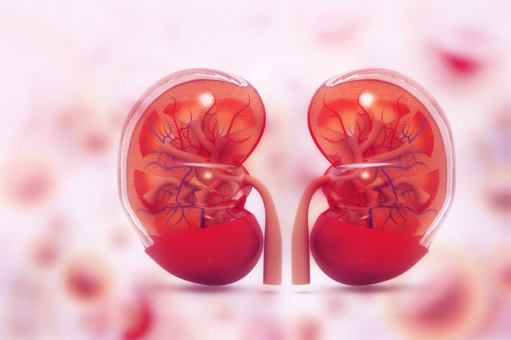
The kidney tubules are the small tubes that collect waste from the blood being filtered at the glomeruli, the network of small blood vessels that compose the kidneys’ filtering system.
“Although Alport disease is initiated in the glomerulus, later stages are characterized by progressive tubulointerstitial fibrosis [scarring] in Alport mouse and in human kidneys. Our detection of sulfatide dysregulation at 28 days, prior to appearance of histopathological [tissue] lesions, suggests that these changes may be involved in very early stages of tubular injury,” the scientists said.


Comments